Figure 5.
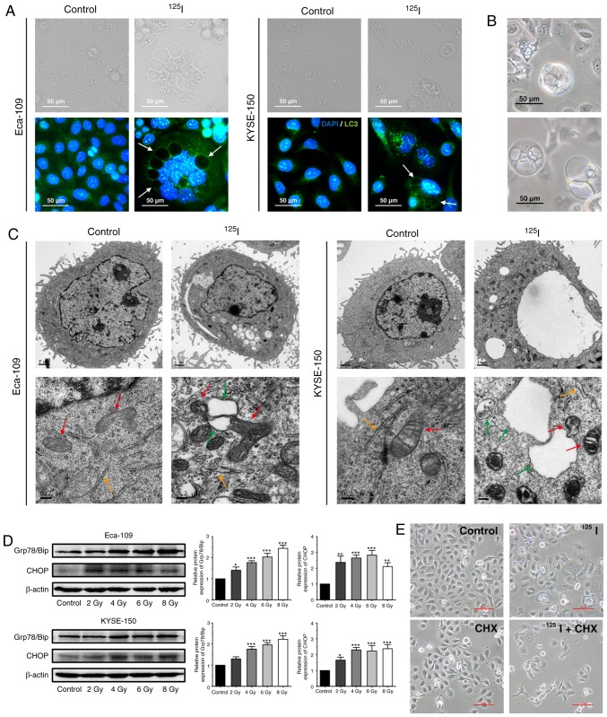
125I seed radiation induces paraptosis in Eca-109 and KYSE-150 cells. (A) Following irradiation, LC3 punctuation (green) was detected under a fluorescence microscope; the nuclei were stained with DAPI (blue). Certain vacuolar membranes appeared negative for anti-LC3 antibody (white arrow). (B) Cell morphology of detached KYSE-150 cells was observed under the light microscope following 4 Gy irradiation. (C) Ultrastructure of Eca-109 and KYSE-150 cells was observed using transmission electron microscopy. Swollen mitochondria (red arrow), endoplasmic reticulum (orange arrow) and vacuoles (green arrow) were observed. (D) Levels of ER stress markers, Grp78/Bip and CHOP, were determined by western blotting. β-actin was used as a loading control. (E) KYSE-150 cells were pretreated with or without CHX, 2 h before 4 Gy irradiation. Morphological changes of KYSE-150 cells were examined under a light microscope. Scale bar, 100 µm. *P<0.05, **P<0.01, ***P<0.001 vs. control group. 125I, Iodine-125; CHX, cycloheximide.

