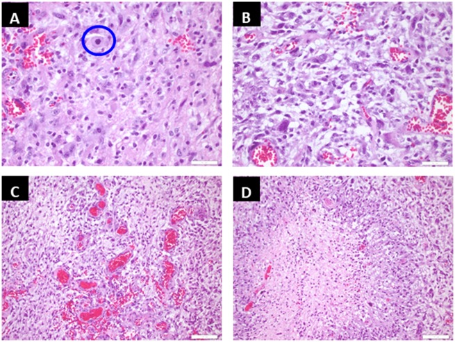
FIGURE 2.
Representative tumor sections from the patient are shown. Hematoxylin and eosin (H&E)-stained section demonstrates neoplastic cells of astrocytic phenotype with an atypical mitosis as shown in the circle (A). Neoplastic cells show prominent pleomorphism (B). There are areas of florid microvascular hyperplasia (C) and geographic necrosis (D), which confirm the diagnosis of glioblastoma.

