
Figure 3.
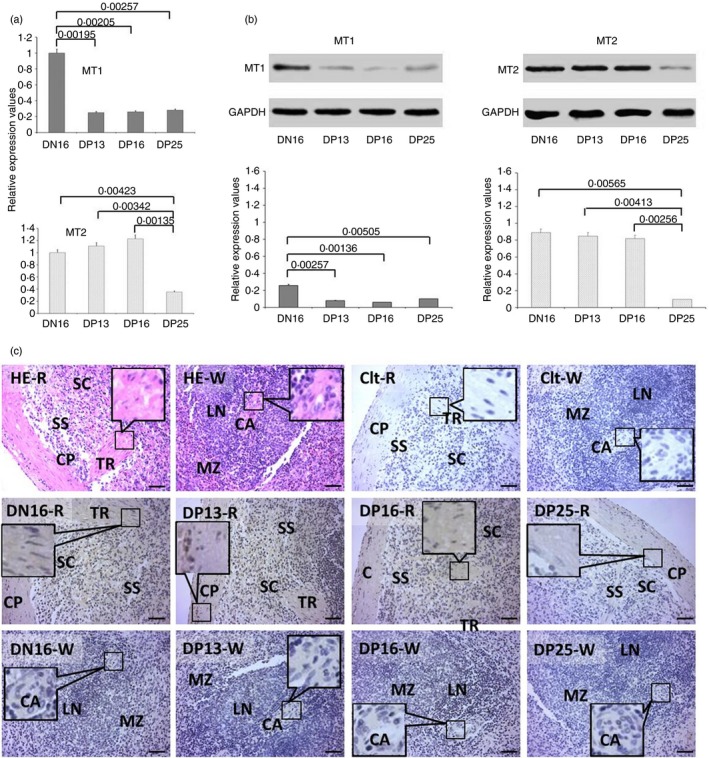
Expression of melatonin receptor 1A (MT1) and melatonin receptor 1B (MT2) in the spleens of non‐pregnant and pregnant ewes. (a) Expression values of MT1 and MT2 mRNA in the spleen. (b) Expression of MT1 and MT2 proteins in the spleen. (c) Representative immunohistochemical localization of MT2 protein in the spleen. The spleen is divided into red pulp (R) and white pulp (W), and surrounded by a thickened capsule (CP). Capsules (CPs) with several trabeculae (TRs) project into the substance of the spleen. Note: HE = stained by haematoxylin and eosin; Ctl = negative control; SS = splenic sinuses; SC = splenic cords; MZ = marginal zone; LN = lymphoid nodule; CA = central arteriole; DN16 = day 16 of the oestrous cycle; DP13 = day 13 of pregnancy; DP16 = day 16 of pregnancy; DP25 = day 25 of pregnancy. Scale bar: 50 µm. P < 0·05 indicates significant difference.