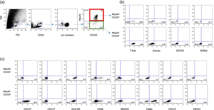
Figure 1.

Phenotypic analysis of intra‐epithelial innate lymphoid cells (ILCs) isolated from normal small intestine biopsies. (a) Gating strategy for CD45+ lineage− [CD3−CD19−CD14−CD16−CD123−CD34−T cell receptor (TCR)γ/δ−TCRα/β−] CD103+NKp44+ and CD103+NKp44− intra‐epithelial ILCs. (b) Intracellular expression of indicated transcription factors in CD103+ NKp44+ (upper panel) and CD103+NKp44− (lower panel) ILCs. (c) Expression of indicated cell surface markers in CD103+NKp44+ cells (upper panel) and CD103+NKp44− cells (lower panel) ILCs. Dot‐plots represent one of three independent experiments.
