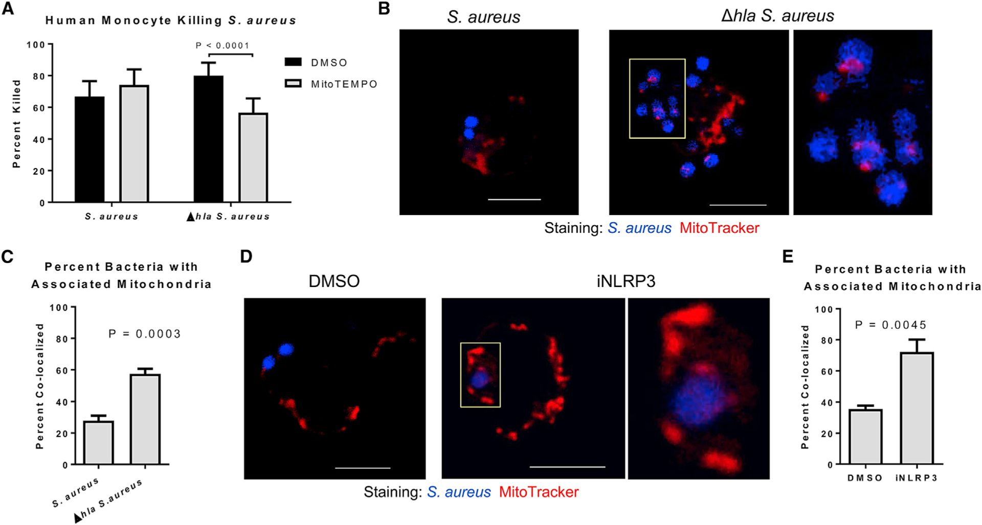
Figure 3. Mitochondria Significantly Contribute to the Killing of Δhla S. aureus.

(A) Percentage of WT or Δhla S. aureus killed following 1-hr incubation with mitoTEMPO- or DMSO-treated (30 min) primary human monocytes.
(B) Confocal images of mitochondria (MitoTracker) and WT or Δhla S. aureus within live primary human monocytes. Scale bar, 5 μm; yellow box indicates the magnified area.
(C) Quantification of the percentage of internalized WT or Δhla S. aureus associated with a mitochondria. n > 100 combined from 3 individual experiments.
(D) Confocal images of mitochondria (MitoTracker) and WT S. aureus within live primary human monocytes treated with MCC950 or DMSO. Scale bar, 5 μm; yellow box indicates the magnified area.
(E) Quantification of the percentage of internalized S. aureus associated with a mitochondria in cells treated with MCC950 or DMSO. n > 50 combined from 3 individual experiments.
Statistical significance (A, C, and E) was determined by Mann-Whitney test. All data are representative of 3 independent experiments. (A) n ≥ 4 replicates per group. Data presented as mean ± SD.
