FIG 5.
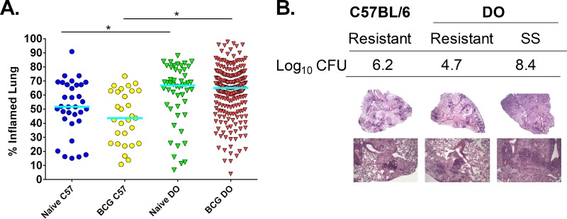
Naive and BCG-vaccinated DO mice challenged with M. tuberculosis exhibit a range of lung inflammation levels and unique lung pathologies. Groups of female B6 and DO mice were vaccinated subcutaneously with 105 M. bovis BCG, and control mice were sham vaccinated with PBS. Mice were challenged by aerosol with ∼45 CFU M. tuberculosis. Animals were euthanized 14 weeks after infection, and lung sections were isolated and prepared for slides with H&E staining. Animals that became moribund before 14 weeks were euthanized, and their lung samples were similarly isolated. H&E-stained slides were scanned, and images were analyzed to assess the percentage of lung tissue with disease involvement compared to the total lung area. (A) Dots represent the percentage of each lung that was inflamed and/or had disease involvement in individual animals, and lines represent the median for the group. Data represent the combined results from five independent experiments similar in design, where the total numbers of animals for each group were 35 naive B6 mice, 60 naive DO mice, 30 BCG-vaccinated B6 mice, and 240 BCG-vaccinated DO mice. (B) Representative images of lung sections prepared for H&E staining that demonstrate various disease pathologies. Lung section images from a BCG-vaccinated B6 mouse with typical disease presentation (resistant), a “resistant” BCG-vaccinated DO mouse that had high lung M. tuberculosis burdens, and a “supersusceptible” mouse with low M. tuberculosis CFU are shown, alongside the log10 lung M. tuberculosis CFU. Images in the top panel used ×2 magnification, and images in the bottom panel used ×10 magnification. *, differences between the groups indicated by the lines were significant (unpaired t test, P < 0.05).

