Figure 5.

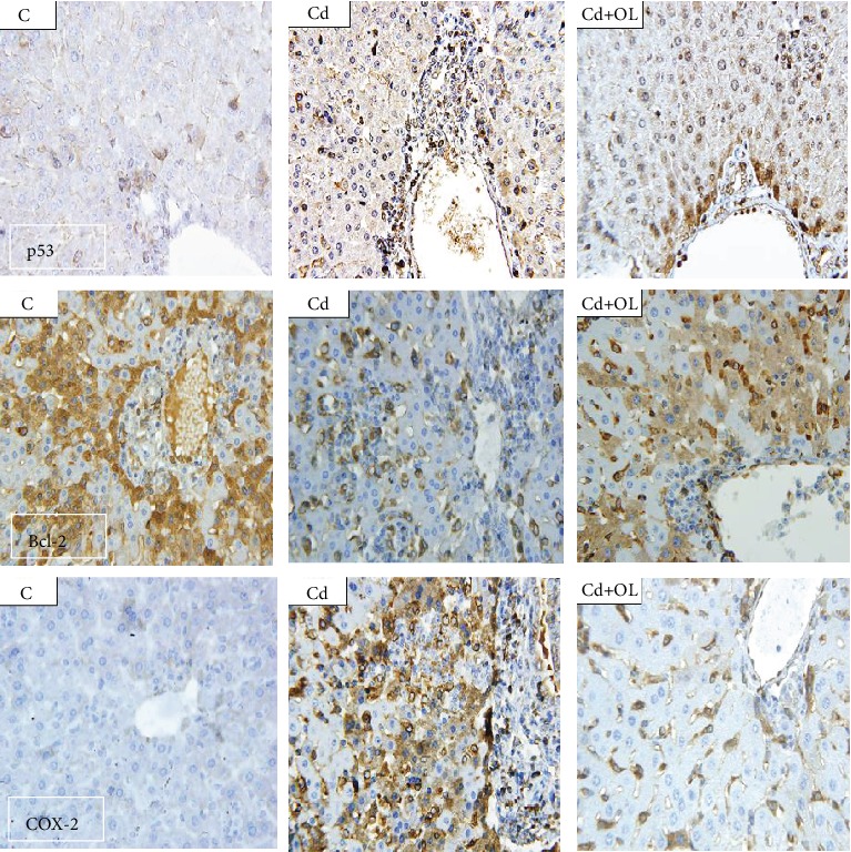
Liver tissue immunohistochemical staining, respectively, with anti-p53, Bcl-2, and COX-2 in the control (C), Cd, and Cd+OL groups (scale bar: 5 μm; magnification: 400x).

Liver tissue immunohistochemical staining, respectively, with anti-p53, Bcl-2, and COX-2 in the control (C), Cd, and Cd+OL groups (scale bar: 5 μm; magnification: 400x).