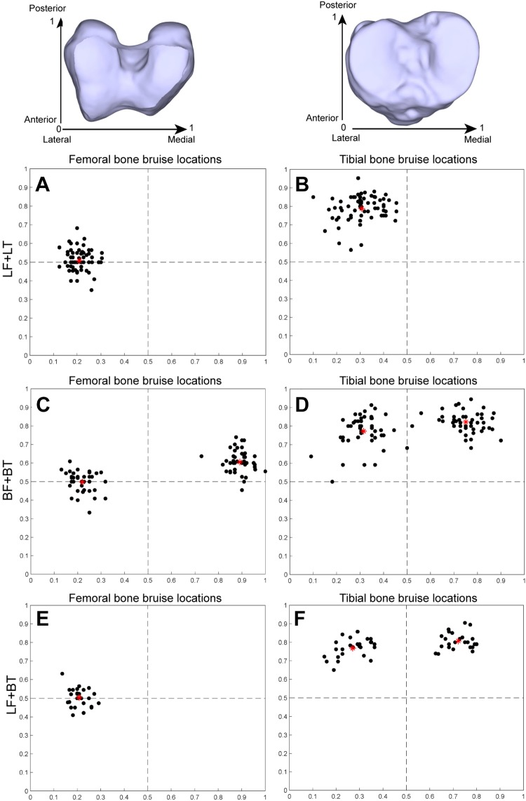
Figure 3.
Bone bruise distribution patterns of the femur and tibia. The dots represent different patients, and the red star in each subfigure represents the mean bone bruise location of those patients. (A) Femoral bone bruise locations of the LF + LT pattern. (B) Tibial bone bruise locations of the LF + LT pattern. (C) Femoral bone bruise locations of the BF + BT pattern. (D) Tibial bone bruise locations of the BF + BT pattern. (E) Femoral bone bruise locations of the LF + BT pattern. (F) Tibial bone bruise locations of the LF + BT pattern. BF, bilateral femur; BT, bilateral tibia; LF, lateral femur; LT, lateral tibia.

