
Figure 3.
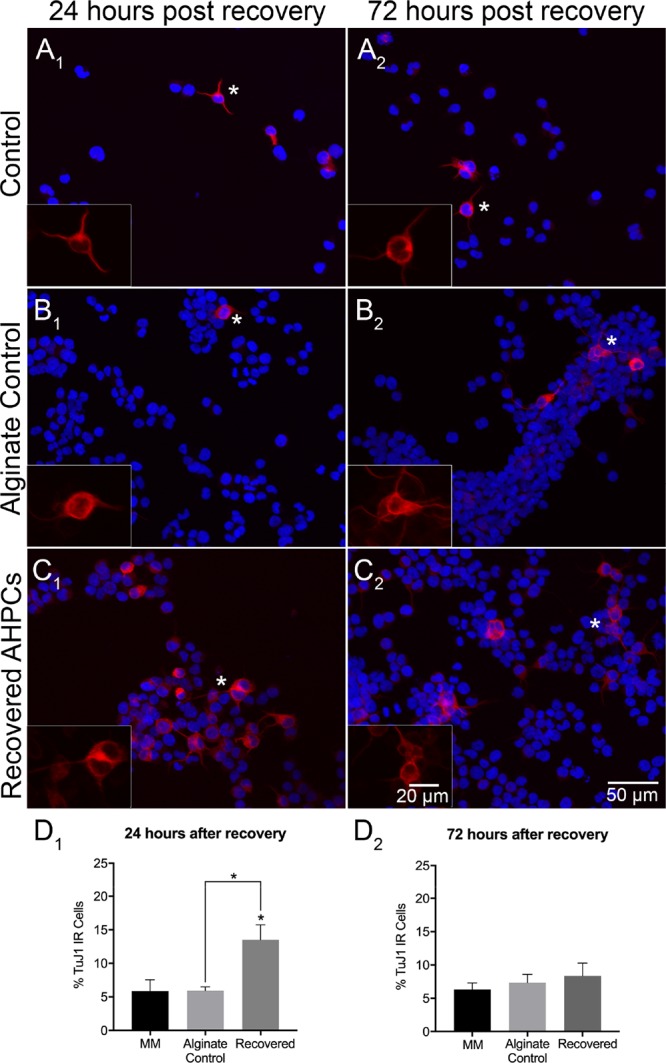
Neuronal differentiation of recovered AHPCs after 4 days of encapsulation. Fluorescence images of AHPCs illustrating immunoreactivity for TuJ1 24 h after recovery (A1–C1) and 72 h after recovery (A2–C2): TuJ1-Cy3 (red) with DAPI staining (blue). AHPCs cultured in MM (A1,A2), alginate control (B1,B2), and recovered from hydrogels (C1,C2). Asterisks indicate the location of higher magnification inset images. Higher magnification images are of the TuJ1-Cy3 channel only. Scale bar = 50 μm (20 μm for insets). Abbreviations: DAPI, 4′,6-diamidino-2-phenylindole. (D1,D2), Quantitative analysis of AHPCs immunoreactive for the TuJ1 antibody. Error bars represent standard error of the mean. N = 6 independent experiments, 5 imaging fields per experiment. *Significantly different at p ≤ 0.05.