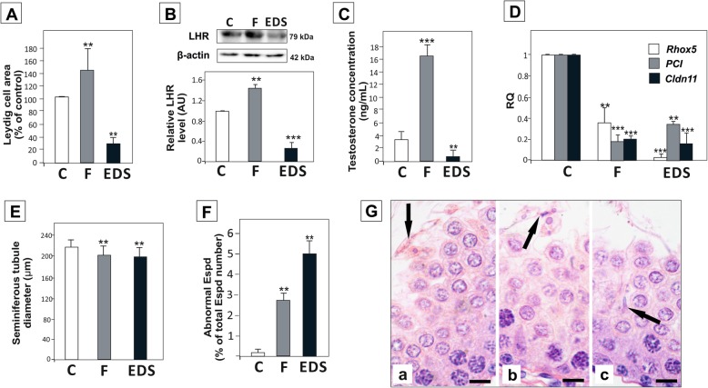
Fig. 1.
Effect of flutamide and EDS-treatment on testicular morphology, plasma testosterone concentration, and the expression of Rhox5, PCI and Cldn11. (A) Area occupied by Leydig cells, (B) LH receptor (LHR) expression in the testis, (C) plasma testosterone concentrations, (D) mRNA expression of Rhox5, PCI and Cldn11, (E) seminiferous tubule diameter, (F) percentage of abnormal or degenerating elongated spermatids (Espd) in seminiferous epithelium of control (C), flutamide (F) and EDS-treated (EDS) rats, and (G) representative images of seminiferous epithelium morphology, showing examples of spermatid degeneration (arrows) in flutamide (Ga) and EDS-treated (Gb, Gc) males; scale bar = 15 μm. For details see Material & methods. Significant differences from control values are denoted as *p < 0.05, **p < 0.01, and ***p < 0.001

