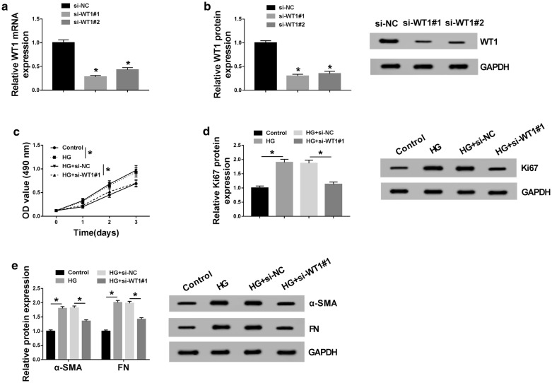
Fig. 3.
HG-induced proliferation and fibrosis in MCs were abolished by WT1 knockdown. WT1 mRNA expression by qRT-PCR (a) and WT1 protein level by western blot (b) in MCs transfected with si-NC, si-WT1#1, and si-WT1#2. MCs were treated with 45 mM of HG or normal medium for 48 h, or transfected with si-NC or si-WT1#1 prior to HG exposure, followed by the measurement of cell proliferation by MTS assay 0, 1, 2 and 3 days after transfection (c), Ki67 expression by western blot after 48 h transfection (d), the levels of α-SMA and FN by western blot 48 h after transfection (e). *P < 0.05

