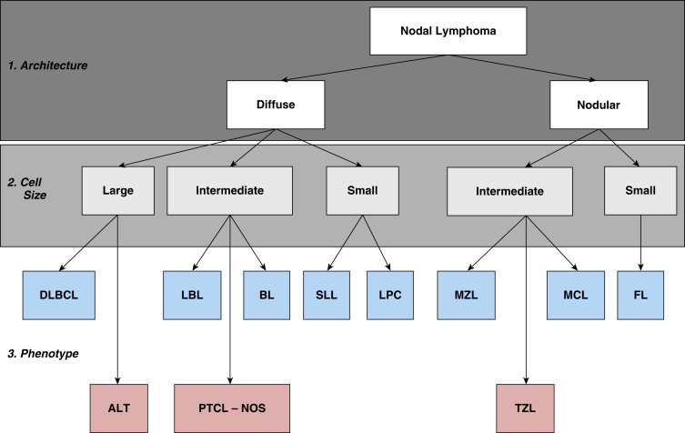
Fig. 33.3.

The histologic approach toward the classification of canine nodal lymphoma. Using excisional lymph node sections, lymphoma is initially divided into diffuse (effacing) or nodular (noneffacing) forms of the disease. Next, using a red blood cell or a small lymphocyte as a guideline, the neoplastic population is divided into large, small, and intermediate forms of the disease. Finally, using knowledge of additional cellular and nuclear features, including mitotic rate and immunophenotype (B cell, blue boxes; T cell, red boxes), a final diagnosis is established. ALT, Anaplastic large cell T-cell lymphoma; BL, Burkitt lymphoma; DLBCL, diffuse large B-cell lymphoma; FL, follicular lymphoma; LBL, lymphoblastic lymphoma; LPC, lymphoplasmacytoid lymphoma; MCL, mantle cell lymphoma; MZL, marginal zone lymphoma; PTCL, NOS, peripheral T-cell lymphoma, not otherwise specified; SLL, small lymphocytic lymphoma; TZL, T-zone lymphoma.
Reproduced and modified with permission from Seelig DM, Avery AC, Ehrhart EJ, Linden MA. The comparative diagnostic features of canine and human lymphoma. Vet Sci. 2016;3(2). Epub 2017/04/25. https://doi.org/10.3390/vetsci3020011. PubMed PMID: 28435836; PubMed Central PMCID: PMCPMC5397114.
