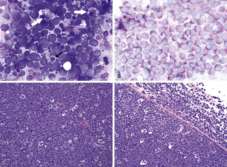
Fig. 33.7.

Lymph nodes from dogs with lymphoma. (A) Fine-needle aspirate. Note the homogeneous population of large lymphoid cells with prominent nucleoli and basophilic cytoplasm. These cells are larger than the neutrophil (black arrow) in the field. Mitotic figures (thin white arrows) and tingible-body macrophages (thick white arrows) also are present. (Wright’s stain, ×60 objective.) (B) Fine-needle aspirate stained for immunoreactivity for CD79a. Note that nearly all of the lymphocytes express CD79a. The diagnosis was B-cell lymphoma. (Alkaline phosphatase/Fast Red, ×60 objective.) (C) Histologic section. Note effacement of normal architecture. The white spaces are macrophages, giving a “starry sky” appearance to the lymph node. (H&E, ×20 objective.) (D) Histologic section. Note the presence of tumor cells outside the capsule of the lymph node. (H&E, ×20 objective.)
