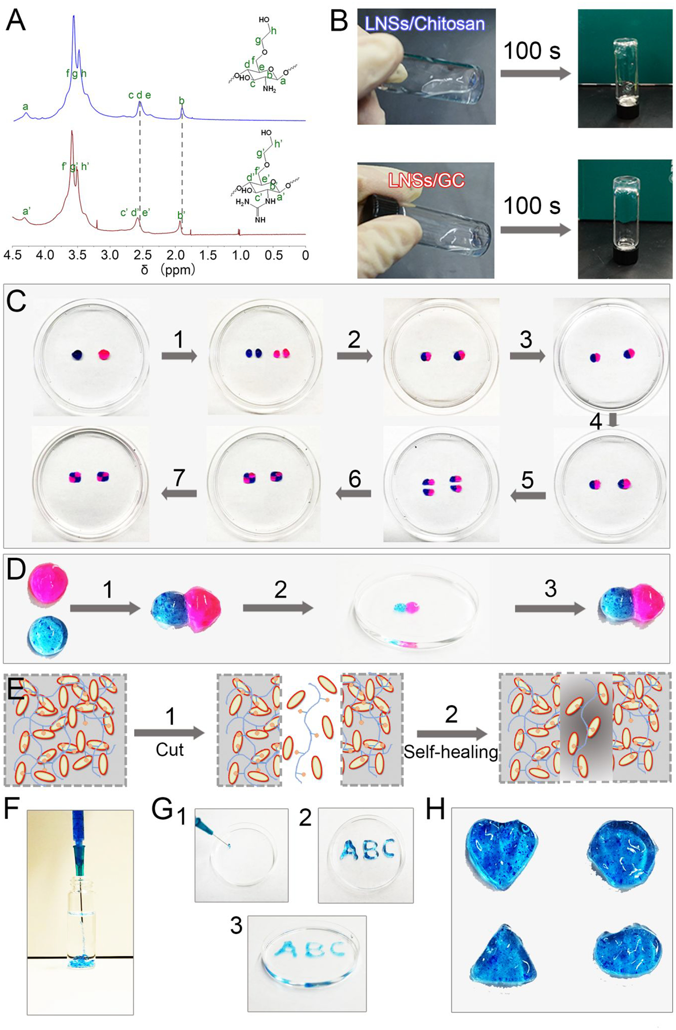
Figure 2.

Physical and chemical properties of supramolecular hydrogels. (A) 1H NMR spectra of the glycol chitosan (blue line) and GC binder (red) in D2O. (B) Images of LNSs with glycol chitosan and LNSs with GC binder. (C) Self-healing processes of supramolecular hydrogels: (1) two disk-shaped hydrogels stained with rhodamine B and methylene blue were cut into equal two pieces; (2) one piece of pink hydrogel and one piece of blue hydrogel were joined together to form two new hydrogels; (3) these two new hydrogels were immersed in PBS; (4) PBS was removed after 3 h; (5) these two new hydrogels were re-cut into equal two pieces; (6) two pieces of pink-blue hydrogels were joined together to form two new hydrogels; (7) these two new hydrogels were immersed in PBS again. (D) Self-healing of two hydrogels after attachment directly: (1) pink hydrogel and blue hydrogel were attached directly; (2) the new hydrogel was immersed into deionized water for 10 min; (3) the hydrogel was transferred from deionized water and maintained its shape. (E) Scheme diagram of the self-healing process. (F) Images of hydrogels injected from 23 G needle into water. (G) “ABC” gel could be formed after injection from 23 G needle. (H) Hydrogels with different shape could be obtained after injection from 23 G needle.
