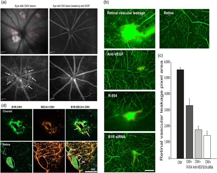
Figure 6.

Effect of anti‐VEGF, R‐954, and B1 receptor siRNA on CNV lesion. CNV is shown as visible hyperfluorescence in the late phase of rat eyes with occult CNV on fluorescein angiography (panel a). The lesion area was more visible in the untreated eyes (left pictures, indicated by red circles and arrows) than in the treated eyes with IVT injection of anti‐VEGF (right pictures). Effects of IVT anti‐VEGF or B1 receptor siRNA and ocular administration of R‐954 on retinal vascular leakage are shown in representative micrographs of the FITC‐dextran (panel b) and quantified by the number of green pixels (panel c). The area of retinal lesions is identified in red in flat‐mounted CNV‐retinas. Data are means ± SEM of values from six rats (five laser spots for each group). * P ≤ .05, significantly different from CNV without treatment. Microphotographs of immunolocalization of B1 receptors (panel d, green) are shown on endothelial cells (panel d, red) in choroid (upper panels) and retina (bottom panels). Scale bar: 200 μm (a), 50 μm (b), 75 μm (d). B1R, B1 receptors
