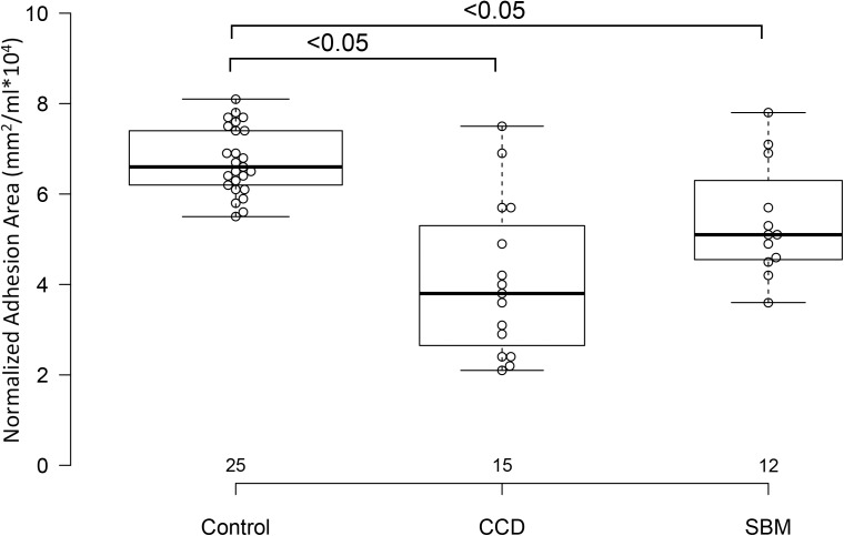
Figure 4. Box and whisker plots of interthalamic adhesion areas normalized to total brain volume for three groups of dogs.
Box and whisker plots of interthalamic adhesion area indexed to total brain volume (TBV) for control dogs, dogs with cognitive dysfunction (CCD) and spontaneous brain microhemorrhage dogs without cognitive dysfunction (SBM).

