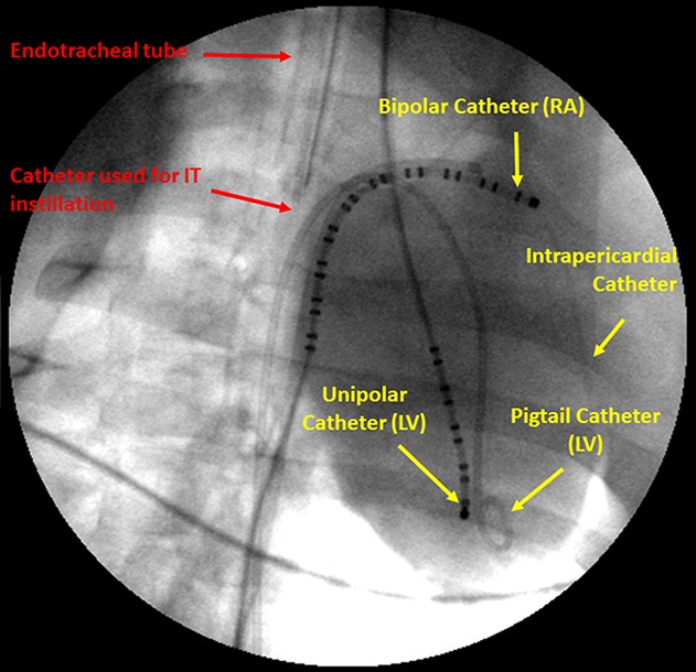
FIGURE 2.

Fluoroscopic image showing the complete set of intratracheal and cardiac catheters. On the left side, red arrows and lettering point to the intratracheal (IT) delivery system, which includes a 5 Fr modified angiography catheter positioned inside the endotracheal tube proximal to the bronchial bifurcation. LV, left ventricle; RA, right atrium. The catheter extends ∼1 cm past the end of the endotracheal tube to allow optimum distribution of the drug bolus. On the right side, yellow arrows and lettering show the RA bipolar catheter and the LV unipolar catheter used for recording electrograms and the LV pigtail catheter to record LV ventricular pressure and compute LV dP/dt as an index of contractility. The RA catheter is also used for pacing. An intrapericardial catheter is used for administering acetylcholine. Published with permission from J Cardiovasc Pharmacol from Marum et al,10 2020.
