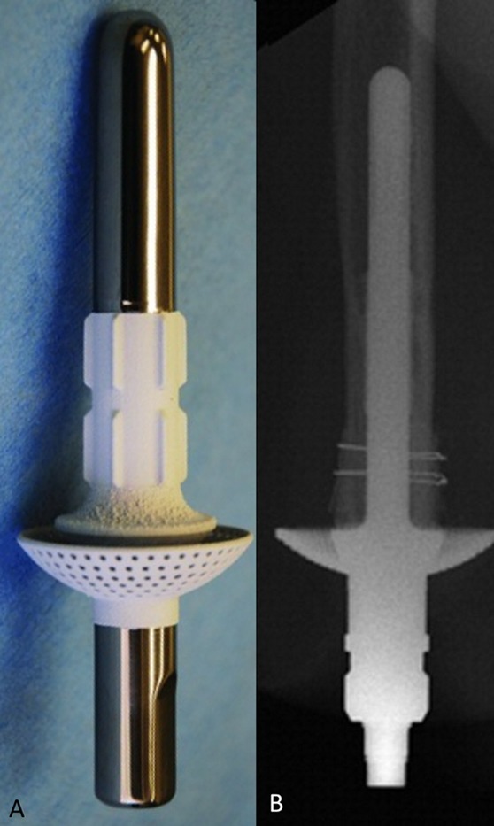
Fig. 6.

ITAP. The implant features a titanium intramedullary stem and a large expansile flanged cap that is coated with hydroxyapatite. The goal of the distal coating was to promote skin adhesion, with the aim of achieving a complete seal against bacterial infiltration. Fig. 6-A The model used in a canine study before the human clinical trial. Note the proximal polished surface with hydroxyapatite coating of the distal portion. (Reproduced from: Fitzpatrick N, Smith TJ, Pendegrass CJ, Yeadon R, Ring M, Goodship AE, Blunn GW. Intraosseous Transcutaneous Amputation Prosthesis [ITAP] for limb salvage in 4 dogs. Vet Surg. 2011 Dec;40[8]:909-25. Epub 2011 Nov 4. © Copyright 2011 by The American College of Veterinary Surgeons. Reproduced with permission.) Fig. 6-B Radiographic view of ITAP in a patient with a humeral amputation. (Reprinted from: J Hand Surg. 35[7], Kang NV, Pendegrass C, Marks L, Blunn G. Osseocutaneous integration of an Intraosseous Transcutaneous Amputation Prosthesis implant used for reconstruction of a transhumeral amputee: case report, 1130-4, 2010. Copyright 2010, with permission from Elsevier.)
