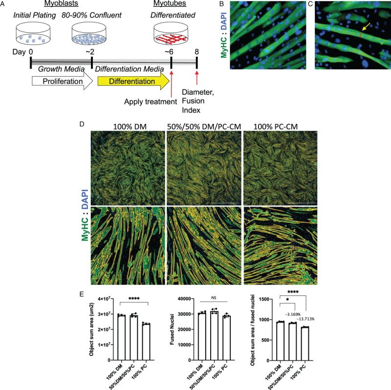
Fig. 2.
Assessing effects on muscle size using C2C12 myotube cultures.

A, Schematic of a typical C2C12 differentiation study in which myoblasts are grown in fetal calf serum-containing growth media, then washed and switched to low growth factor containing differentiation media (DM) for 4 days, then incubated with test factors for 48 h, then washed, fixed, and measured on day 8. B, Myotubes are visualized by immunofluorescence with antimyosin heavy chain antibody (green) and DAPI to visualize nuclei (blue). C, Manual measurements on calibrated micrografts can be done manually using ImageJ. D, Alternatively, wells can be scanned on digital scanning microscopes (Lionheart was used here), and E, automated image analysis used to determine total area covered by myotubes (object sum area), the number of nuclei within myotubes (fused nuclei), and the ratio. Here 4-day-old myotubes were incubated for 48 h in 100% DM or 50% DM with 50% pancreatic cancer cell conditioned media (PC-CM) or 100% PC-CM. Automated imaging demonstrates a reduction in overall green area, a non-significant reduction in fused nuclei, and reductions in the ratio, indicating smaller myotube cell body size per nucleus or atrophy. ∗P < 0.05; ∗∗∗∗P < 0.0001. These studies were done using four replicate wells per condition.
