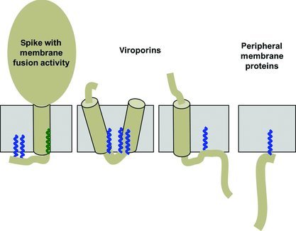
Figure 1.

Classification of S‐acylated proteins from viruses
The figure shows the membrane topology of S‐acylated viral proteins. The (presumably) α‐helical transmembrane region is depicted as cylinder embedded within a membrane (grey). Fatty acids linked to cysteine residues are shown as zigzag line. Acylation sites located at the end of the cytoplasmic part of the transmembrane region of viral spike proteins often contain stearic acid (green), acylation sites within the cytoplasmic tail contain palmitic acid (blue).
