
Figure 5.
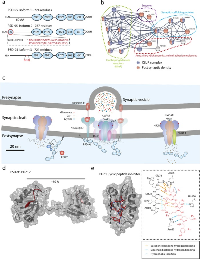
a) Domain organization of PSD‐95 (Uniprot: P78352) and its splice variant derived isoforms. b) A protein interaction network (STRING) showing a selection of 20 proteins (highest confidence score) interacting with PSD‐95 shows high interconnectivity between the different proteins in their respective groups. Made using STRING database information, analysis, and visualization tools. c) Graphical illustration of selected membrane protein/PSD‐95 PDZ interactions in the postsynaptic density. d) Structure of PSD‐95 PDZ1‐2 in its double Cypin (NH2‐QVVPFSSSV‐COOH) occupied state shows parallel orientation of the PDZ1 and PDZ2 binding pocket (PDB: 2KA9). e) Structure (left) and hydrogen bonding network (right) of cyclic lactam‐containing peptide (NH2‐YK‐c[KTE(βA)]‐V‐COOH) insertion into PDZ1 of PSD‐95 displaying additional hydrogen bonds compared to canonical type II ligand insertion (PDB: 1RGR).