
Figure 4.
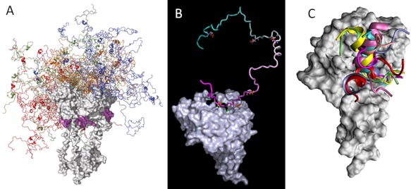
Fuzziness of protein structures and complexes. A: Fuzzy structure of a hybrid protein (p53 tetramer) that contains structured DNA‐binding and tetramerization domains (gray space‐filling models) and a disordered transactivator domain (shown as an ensemble of 20 conformations in different colors for each molecule in the tetramer). Figure is modified from Ref. 253 with permission. B: The NMR structure of a fuzzy complex between the cyclin‐dependent kinase inhibitor Sic1 [depicted as a ribbon with color‐coding from cyan (N‐terminus) to magenta (C‐terminus)] and the ubiquitin ligase Cdc4 (depicted as space‐filling gray model). At any given moment, only one out of the nine phosphorylated sites of Sic1 interacts with a single binding site in Cdc4, generating a highly dynamic conformational ensemble of a complex described within the frames of the “polyelectrostatic” model.254, 255 C. Fuzzy complex of the negative regulatory domain (NRD) of p53 with dimeric S100B(ββ). According to the extensive all‐atom explicit solvent simulations, NRD of p53 remains highly dynamic in the S100B(ββ)‐bound state.256