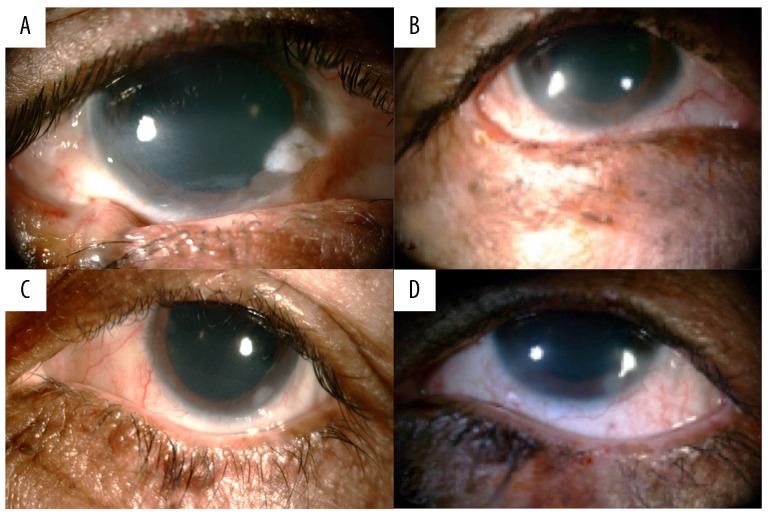
Figure 2.
Four images from Case 2 taken 2 years apart. (A) Right eye before treatment with basal cell carcinoma involving the lateral third of the lower eyelid and extending to the right lateral canthus. (B) Right eye after surgical excision and 21 months of therapy. (C) Left eye before treatment, showing the cornea with the limbal 360-degree involvement by an irregular hazy white growth which was identified as fibrosis with no malignant cells on biopsy. (D) Left eye after surgical excision and 21 months of therapy.

