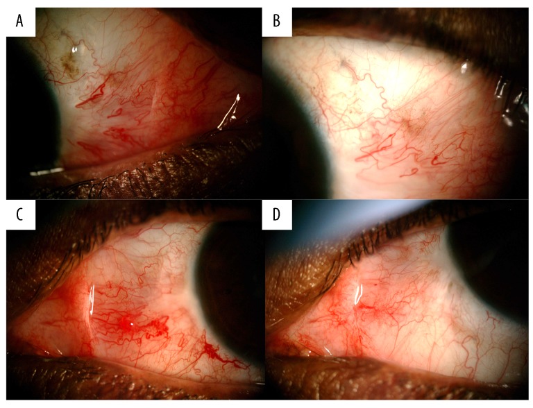
Figure 3.
Four images of Case 3 taken 2 years apart. (A) Post-surgical excision on the right eye, showing focal squamous cell carcinomas in situ at 03: 00 limbal area. (B) Right eye after 18 months of therapy. (C) Post-surgical excision on left eye showing focal epithelial dysplasia at limbal conjunctiva with irregular vascularization. (D) Left eye after 18 months of therapy.

