
Figure 1.
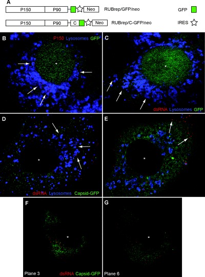
Immunofluorescence and confocal microscopy localization of RUB replication components in replicon‐transfected cells (Vero cells, 2 or 4 days post transfection). Single optical sections are shown. An asterisk marks the centre of the cell nucleus. A. Schematic diagrams of the two replicons used in this study, RUBrep/GFP/neo and RUBrep/C‐GFP/neo. ORFs are represented by boxes and untranslated regions by lines. B. Localization of P150 non‐structural protein (red) and lysosomes (blue) in cells transfected with RUBrep/GFP/neo. Lysosomes were labelled with anti‐LAMP‐1 antibodies. Arrows indicate P150 signal within lysosomes. With this construct, GFP localizes in both the cell nucleus (*) and the cytoplasm. C. Localization of dsRNA (antibody labelled in red) and lysosomes (blue) in cells transfected with RUBrep/GFP/neo. Arrows indicate dsRNA signal within lysosomes. D and E. Localization of dsRNA (red) and lysosomes (blue) in cells transfected with RUBrep/C‐GFP/neo. In D, arrows indicate dsRNA signal within lysosomes while in E, arrows indicate localization of dsRNA signal on cell periphery. With this construct, the C‐GFP fusion accumulates in the cytoplasm. F and G. Two confocal single sections of the same cell transfected with RUBrep/C‐GFP/neo and stained for dsRNA (red) showing dsRNA concentrating in the perinuclear region in the central section (F) or on the cell periphery in the lower section (G).