
Figure 2.
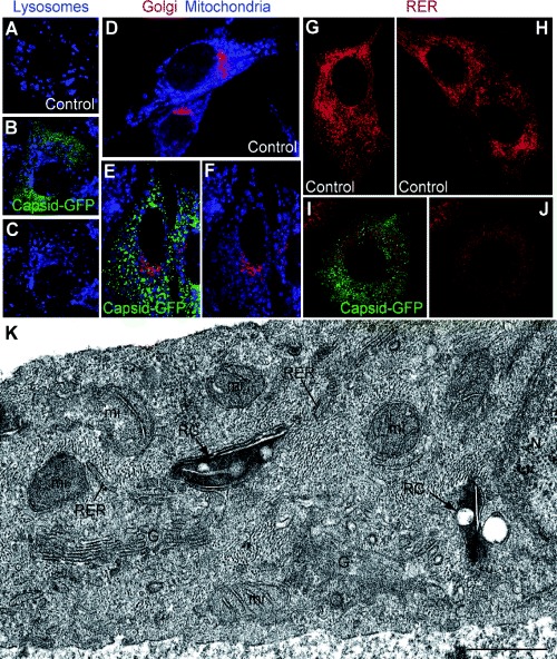
Organelle distribution and ultrastructure in replicon‐transfected cells (control non‐transfected BHK‐21 cells, BHK‐21 cells stably transfected with RUBrep/C‐GFP/neo, and Vero cells transiently transfected with this replicon at 2 days post transfection). Single optical sections are shown. Vero cells are shown in A–C while the rest of fields are of BHK‐21 cells. The cells were stained for lysosomes (blue) (A–C), Golgi (red) and mitochondria (visualized with Mitotracker blue) (D–F) and RER (red) (G–J). Panels B/C, E/F and I/J show the same field; in C, F and J, the green signal has been removed to allow better resolution of the organelle staining pattern. (K) EM of perinuclear areas of cells transfected with RUBrep/C‐GFP/neo. Dense, modified lysosome‐like organelles or CPVs (‘RC’ for replication complex) locally recruit mitochondria (mi) in the proximity of the cell nucleus (N) and are surrounded by RER, and Golgi stacks (G). Bar: 1 μm.