
Figure 4.
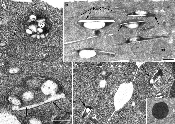
Ultrastructural analysis of CPVs in replicon‐transfected and RUB‐infected cells by conventional processing for thin‐section and EM (BHK‐21 cells transfected with RUBrep/C‐GFP/neo after drug selection). A and B. Conventional EM shows two views of electron‐dense CPVs in perinuclear areas of replicon‐transfected cells. Internal vesicles (V) and straight elements (arrows) are denoted as are locally recruited organelles, RER and mitochondria (mi). The CPVs in B appear to be side views of the CPVs while in A, a frontal view of the CPV is shown. C and D. CPVs in RUB‐infected BHK cells 24 (C) and 48 (D) hours post infection. C is a higher magnification while D is a lower magnification. CPVs in infected cells contain both vesicles and straight elements (arrows). Inset in D shows a characteristic dense lysosome in a control cell. V, vesicles; N, nucleus. Bars: 200 nm in A–C; 0.5 μm in D.