
Figure 4.
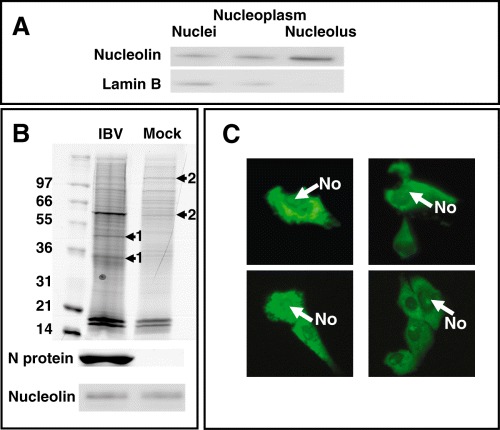
A. Western blot analysis of consecutive stages in the preparation of nucleolar extracts. Nucleolin is used as a marker for the nucleolus and Lamin B as a maker of the nucleus and nucleoplasm. B. Top image, Coomassie stained acrylamide gel of nucleolar extracts from IBV and mock‐infected cells separated on a 10% NuPage Bis‐Tris precast polyacrylamide gel (Invitrogen) in MOPs running buffer. Arrows indicated both novel protein species (1) and reduced cellular proteins (2) in the nucleolar extracts from infected and mock‐infected cells respectively. Below are shown Western blots for N protein and nucleolin. C. Live cell imaging of cells expressing EGFP‐IBV N protein. The nucleolus is indicated (No).