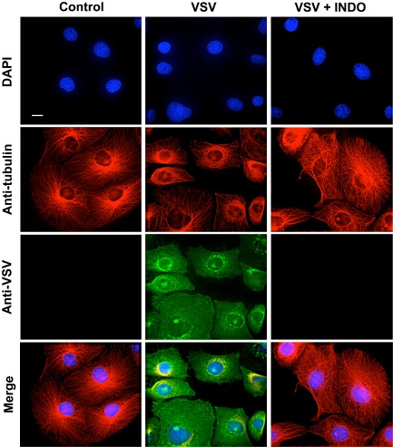
Figure 2.

Effect of indomethacin on VSV protein expression and intracellular localization. MA104 cells mock infected or infected with VSV (10 PFU per cell) were treated with 400 μM INDO after the 1 h adsorption period and, at 6 h p.i., viral proteins were visualized by immunofluorescence microscopy using anti‐VSV (green) and anti‐α‐tubulin (red) antibodies. Nuclei are stained with 4′,6‐diamidino‐2‐phenylindole (DAPI) (blue). The overlay of the three fluorochromes is shown (Merge). Images were collected and deconvolved with DeltaVision microscope and software. Bar = 15 μm.
