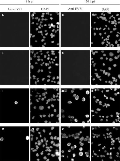
Figure 2.

Immunofluorescence microscopy of uninfected and EV71‐infected RD cells at 8 and 20 h p.i. Infected cells were detected by anti‐EV71 monoclonal antibody, while the cell nuclei were displayed by DAPI staining. A–D. Uninfected RD cells. E–H. RD cells mock‐infected with inactivated EV71 strain MS/7423/87. I–L. RD cells infected with EV71 strain MS/7423/87. M–P. RD cells infected with EV71 strain 5865/Sin/000009.
