
Figure 5.
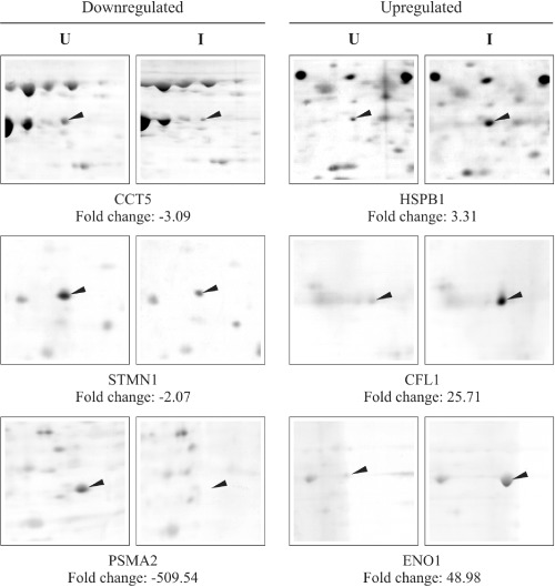
Identification of six differentially expressed proteins by two‐dimensional proteomic analysis of uninfected (U) and EV71 MS/7423/87 strain‐infected (I) RD cell samples at 8 and 20 h p.i. The arrowheads indicate the protein spots identified as differentially regulated at greater than twofold changes (P < 0.05) using Delta2D analysis.