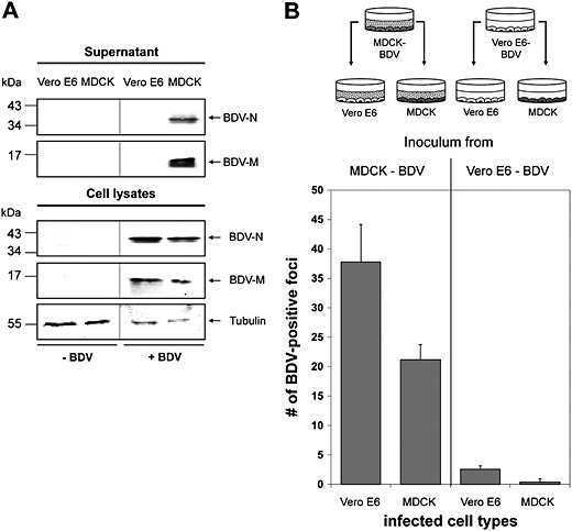
Figure 1.

Cell type‐dependent release of free BDV particles. A. Persistently BDV‐infected MDCK and Vero E6 cells were grown to confluency. Uninfected cells served as controls. After further incubation for 4 days, BDV was isolated from the cell culture supernatant and subjected to SDS‐PAGE followed by immunoblotting using BDV‐N‐specific and BDV‐M‐specific antisera. Tubulin present in the cell lysates was used as a loading control and detected using an anti‐tubulin antibody. B. Non‐infected MDCK and non‐infected Vero E6 cells were grown on glass coverslips and incubated for 48 h with pre‐cleared cell culture supernatants from either persistently BDV‐infected MDCK or persistently‐BDV infected Vero E6 cells as depicted. After further incubation for 48 h, each cell culture was fixed, incubated with a BDV‐N specific antibody and stained using an HRP‐coupled secondary antibody and TrueBlue as substrate. BDV‐infected cell foci were counted in three randomly chosen areas.
