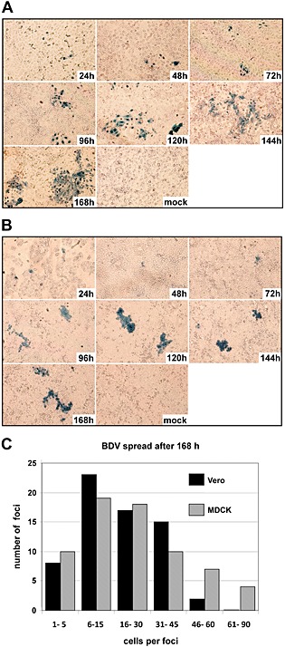
Figure 2.

Kinetics of BDV spread in MDCK and Vero E6 cell cultures. Non‐infected MDCK cells (A) and Vero E6 cells (B) were mixed with homologous, infected cells at a ratio of 1 infected to 400 uninfected cells and were grown on glass coverslips. At the indicated time points, individual coverslips were removed; the cells were fixed, permeabilized, incubated with a BDV‐N‐specific antibody and stained using an HRP‐coupled secondary antibody and TrueBlue substrate to visualize BDV‐infected cells. The non‐infected control cells (mock) were analysed at 168 h after seeding on coverslips. (C) Quantification of virus spread. MDCK and Vero E6 cells were infected as described for (A) and (B). After 168 h, the number of immunostained cells per focus was counted and foci were categorized depending on the number of BDV‐positive cells per focus.
