Figure 5.

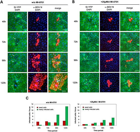
Cell‐to‐cell spread of BDV in MDCK cells in the presence or absence of GP cleavage.
Non‐infected MDCK cells persistently expressing SI‐YFP were mixed with BDV‐infected MDCK cells (seed cells) at a ratio of 100:1 and grown on glass coverslips in the absence (A) and in the presence (B) of 25 μM of MI‐701 respectively. The culture medium was exchanged with fresh medium with or without inhibitor every 24 h. At the indicated time points, individual coverslips were removed and cells were fixed, permeabilized, incubated with a BDV‐N‐specific antibody and stained using a Cy5‐conjugated secondary antibody (red). Nuclei were stained with DAPI (blue). Arrows indicate the position of seed cells of which the nuclei were stained with DAPI. Scale bars, 50 µm. Virus spread inhibition was quantified by using two independent experiments.
C. The MI‐0701 mediated reduction of BDV spread was determined by comparison of the numbers of BDV infected cells, which are double labelled for fluorescence label with Cy5 and YFP (yellow fluorescence protein) at different time points (48, 72, 96 and 120 h).
