Figure 6.

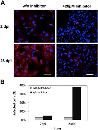
Cell‐to‐cell spread of BDV in primary rat astrocytes in the presence or absence of GP cleavage.
A. Non‐infected primary cortical rat astrocytes were mixed with BDV‐infected primary cortical rat astrocytes at a ratio of 100:1 and co‐cultivated for 23 days in the presence or absence of 20 μM of MI‐0701. At the indicated time points, individual samples were fixed, permeabilized, incubated with an antibody specific for BDV‐N and stained using a Cy5‐conjugated secondary antibody (red). Nuclei were visualized by staining with DAPI. Scale bars, 50 µm.
B. Quantification of total number of infected astrocytes.
