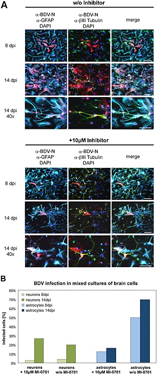
Figure 7.

Influence of GP cleavage on the cell‐to‐cell spread of BDV in mixed cultures of brain cells.
A. Cultures of primary rat cortical astrocytes and neurons were infected with BDV and cultured for 14 days in the presence or absence of 10 μM of MI‐0701. At 8 and 14 dpi, individual samples were fixed and permeabilized. BDV was detected using a BDV‐N‐specific antibody and a Texas Red‐conjugated secondary antibody (red). Astrocytes were stained using an α‐GFAP antiserum and a Cy5‐conjugated secondary antibody (cyan). Neurons were detected using an α‐βIII Tubulin antiserum and an FITC‐conjugated secondary antibody (green). Nuclei were stained with DAPI. Scale bars, 50 µm.
B. Quantification of BDV‐infected astrocytes and neurons for an experiment conducted as described in (A).
