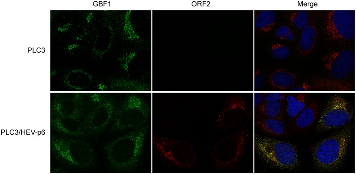
Figure 6.

Intracellular distribution of GBF1 in hepatitis E virus (HEV)‐replicating and non‐replicating cells. PLC3 cells were electroporated with water or with the full‐length infectious p6 strain RNA. At 3 days p.e., cells were fixed, permeabilized, and processed for double‐label immunofluorescence for GBF1 (red) and ORF2 (green). Nuclei are in blue. Representative confocal images are shown together with the merge image
