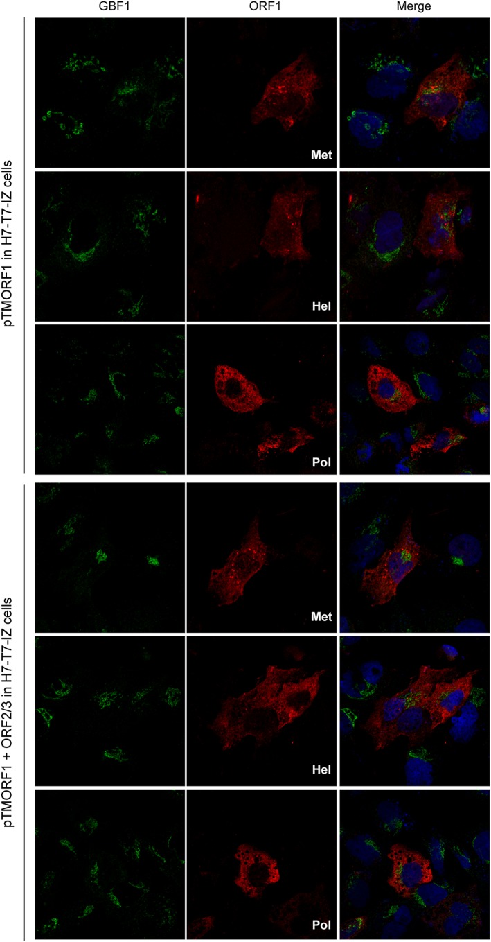
Figure 7.

Intracellular distribution of GBF1 in hepatitis E virus ORF1‐expressing cells. H7T7IZ cells were transfected with pTM‐ORF1 or in combination with pTM‐ORF2/3. Twenty four hours post‐transfection, cells were fixed, permeabilized, and processed for double‐label immunofluorescence for GBF1 (green) and ORF1 methyltransferase , helicase, or polymerase domain (red). Nuclei are in blue. Representative confocal images are shown together with the merge image
