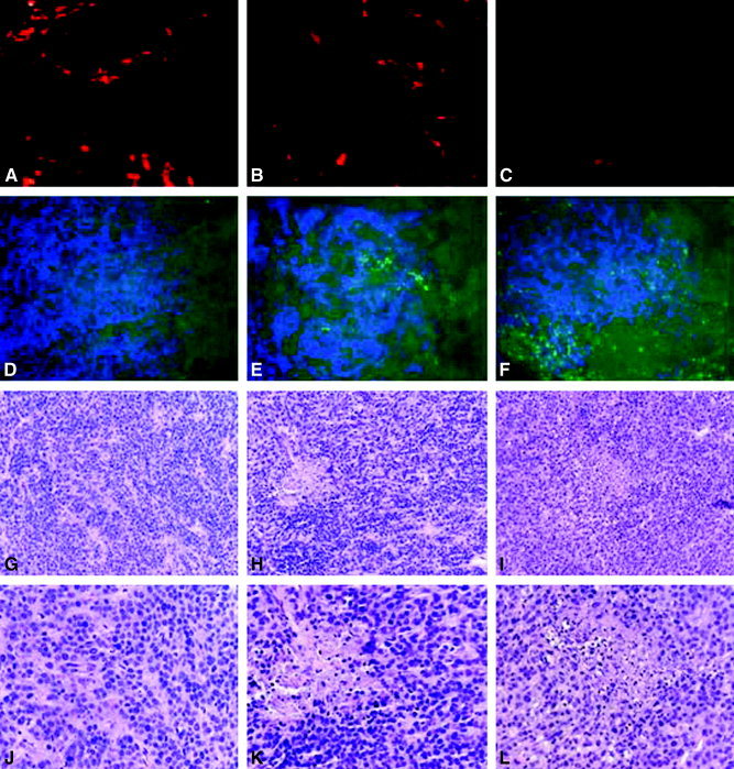
Figure 4.

Apoptosis and histochemical analysis. Residual tumors from endostatin‐treated groups and from NGR sequence‐modified endostatin (NGR‐endostatin)‐treated groups were resected 1 day after the completion of treatment. A–C show vessel density, as determined by phycoerythrin‐labeled, anti‐CD31 antibody staining; D–F show terminal deoxyuridine triphosphate nick‐end labeling assay (green) with 4,6‐diamidino‐2‐phenylindole (blue); G–L show hematoxylin and eosin staining. A, D, G, and J are control sections; B, E, H, and K are endostatin‐treated tumor sections; C, F, I, and L are NGR‐endostatin‐treated tumor sections. Original magnification ×100 (A–I); ×400 (J–L).
