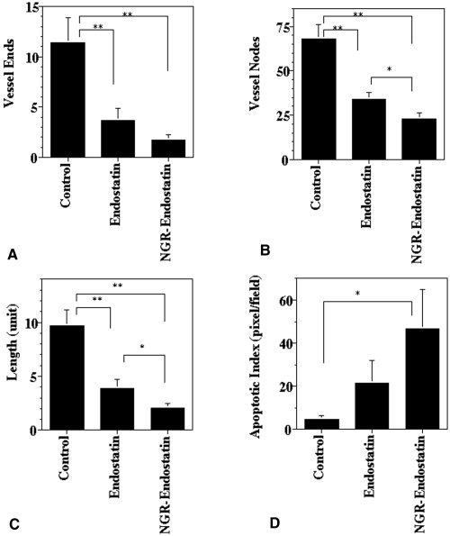
Figure 5.

Quantification of antiangiogenic and apoptotic effect. Seven to 10 frames of tumor sections that were stained with antimouse CD31‐phycoerythrin were captured per sample and then analyzed for microvessel density. (A) Blood vessel ends. (B) Branch points (vessel nodes). (C) Vessel length. (D) Quantification of apoptotic cells (pixel = cells) showed a marked increase in NGR sequence‐modified endostatin (NGR‐endostatin)‐treated animals. Statistical significance was determined with a Student t test. Single asterisk: P < 0.05; double asterisks: P < 0.01. Error bars indicate the standard error.
