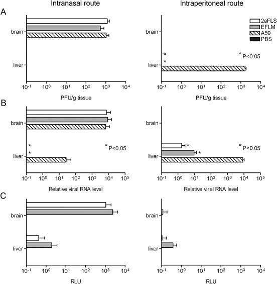
Figure 2.

Replication of MHV‐EFLM and MHV‐2aFLS in vivo. The 6‐ to 8‐week‐old female BALB/c mice were inoculated either intranasally or intraperitoneally with PBS or with 106 TCID50 of wild‐type MHV‐A59, MHV‐EFLM or MHV‐2aFLS. At 5 days post inoculation, all mice were sacrificed and brains and livers were collected. Homogenates were prepared as described in the Experimental procedures. A. Viral infectivity in the homogenates was determined by performing a plaque assay on LR7 cells. The viral titres are expressed as plaque‐forming units per gram (PFU g−1) tissue. B. The amounts of viral genomic RNA relative to total RNA, isolated from the liver and brain homogenates, were determined by quantitative Taqman RT‐PCR. C. Luciferase activities (in RLU) in each of the homogenates were determined by using a luminometer as described above. Standard deviations (n = 4) are indicated in all graphs.
