Figure 5.

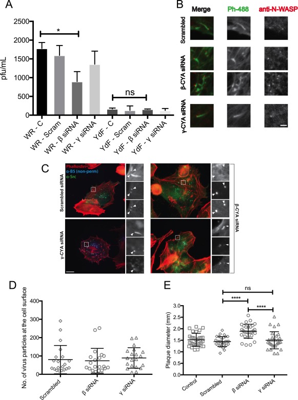
Effect of β‐CYA depletion on VACV signalling and spread. (a) HeLa cells were treated with the specified siRNA and then infected with the specified VACV (WR strain or VACV‐A36YdF, depicted as YdF), at an MOI of 0.1. Supernatants were collected at 16 hpi and used to perform plaque assays on BSC‐1 cells to estimate viral titre (“ns”: p > .05, “*”: p ≤ .05, n = 3). (b) VACV‐infected cells under CYA knockdown were fixed 8 hpi and stained for N‐WASP (red) and phalloidin (green). Scale bar is 1 μm. (c) VACV‐infected cells under CYA knockdown were fixed 8 hpi and stained for virus envelope protein B5 (nonpermeablized, blue), phallodin (red), and anti‐Src (green). Scale bar is 10 μm. (d) HeLa cells were treated with CYA siRNA and infected with VACV‐WR at an MOI > 3. Cells were fixed 8 hpi, followed by staining with anti‐B5 prior to permeabilization. The number of virus particles at the cell surface was counted in each case (n = 10 cells each, in 2 experimental replicates). (e) GBM cells were treated with the specified CYA siRNA and infected with VACV‐WR at an MOI of 0.1. Cells were fixed 3 dpi and stained to measure plaque diameters. (“****”: p ≤ .0001, n = 30 plaques each, with 2 experimental replicates). [Color figure can be viewed at http://wileyonlinelibrary.com]
