
Figure 3.
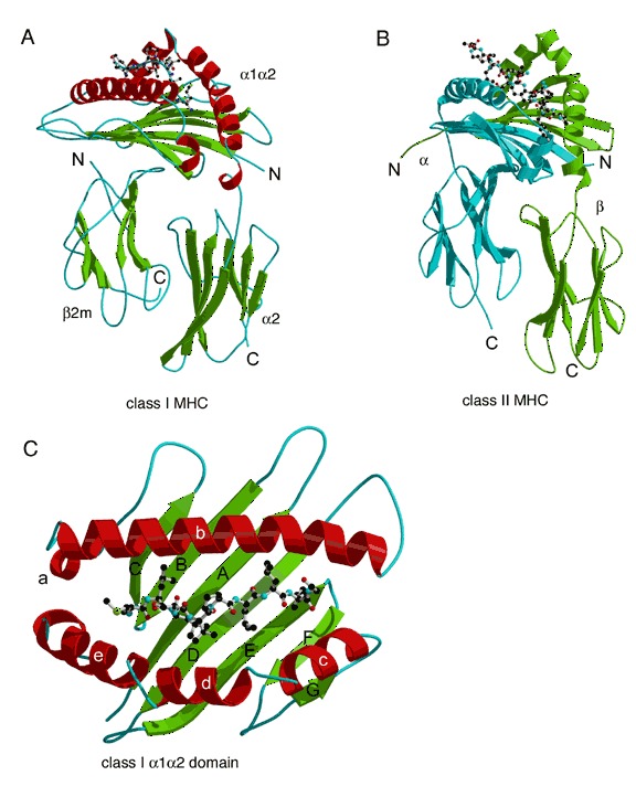
MHC structures. (A) Class I MHC HLA‐A2 complex (PDB entry 2clr) and (B) class II MHC complex HLA‐DR1 (PDB entry 1dlh), each with an antigenic peptide. The α and β chains in (B) are colored blue and green respectively. The peptide is shown as a ball‐and‐stick model. (C) Close‐up view of the α1α2 peptide‐binding domain of HLA‐A2 (PDB entry 2clr).