
Figure 5.
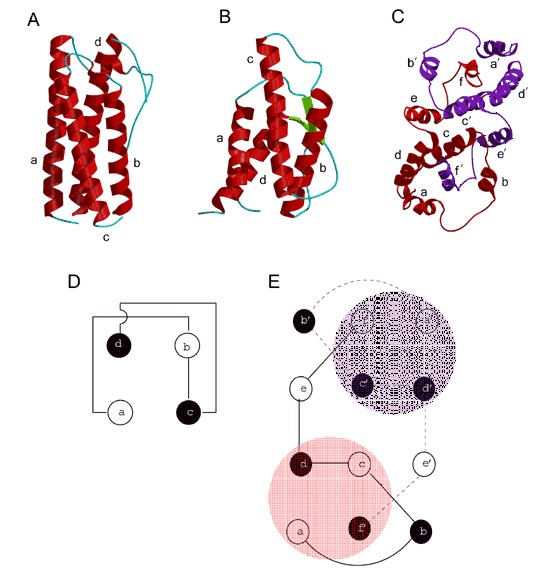
Long and short‐chain cytokines. The structure of (A) a long‐chain helical cytokine, GCSF (PDB entry 1rhg), (B) a short‐chain helical cytokine, IL‐4 (PDB entry 1rcb), and (C) interferon‐γ (PDB entry 1rfb). The two monomers of interferon‐γ are shown in red and purple. (D) Connectivity between helices in four‐helix bundle cytokines. The up helices are drawn in white and the down helices are in black. (E) Connectivity between helices of IFN‐γ. The block shaded regions correspond to the four‐helix bundles.