
Figure 6.
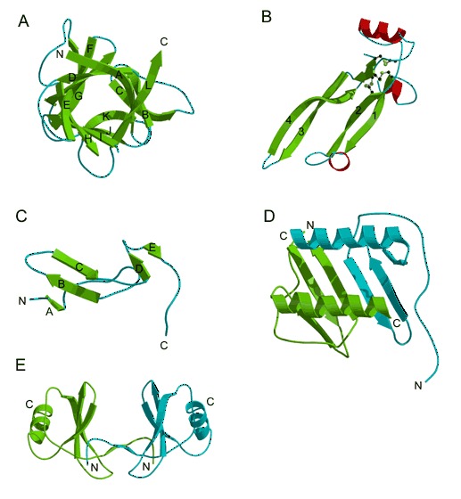
Cytokines and chemokines. (A) Human IL‐1β, a member of the β‐trefoil fold. (PDB entry 1hib). (B) Human transforming growth factor‐β2 (PDB entry 2tgi). The four strands that define the cysteine‐knot fold are shown in green (Anderson et al., 1978). The six knotted cysteines are shown in ball‐and‐stick model (with yellow sulfur atoms). (C) Murine EGF (PDB entry 1epj). (D) CXC chemokine (PDB entry 1il8), (E) CC chemokine (PDB entry 1rto).