
Figure 8.
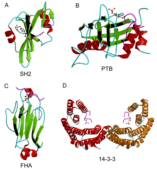
Folds that bind phosphopeptides. The phosphopeptides are shown as magenta colored worms. The phosphorylated amino acid is represented by a ball‐and‐stick model. (A) The SH2 domain from v‐src tyrosine kinase bound to a five‐residue phosphotyrosine peptide (PDB entry 1sha). (B) PTB domain from shc complexed with a twelve‐residue phosphotyrosine peptide (PDB entry 1shc). (C) FHA domain from protein kinase RAD53 complexed to a twelve‐residue phosphothreonine peptide (PDB entry 1g6g). (D) Homodimer of 14‐3‐3 protein ζ bound to eight‐residue phosphoserine peptides (PDB entry 1qja).