
Figure 9.
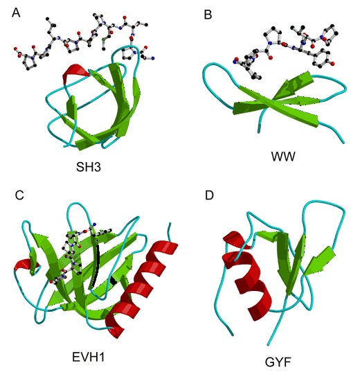
Folds that bind to polyproline peptides. Bound polyproline peptides are represented by ball‐and‐stick models. (A) An SH3 domain from the Abl tyrosine kinase complexed with the ten‐residue synthetic peptide 3Bp‐1 (PDB entry 1abo). (B) A WW domain from dystrophin in complex with a seven‐residue β‐dystroglycan peptide (PDB entry 1eg4). (C) An EVH1 domain from Enabled, bound to the Acta peptide (PDB entry 1evh). (D) GYF domain from CD2Bp2 (PDB entry 1gyf).