
Figure 10.
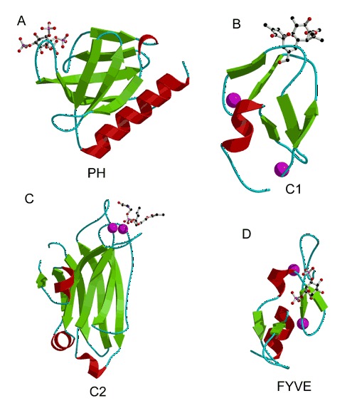
Phospholipid‐binding domains. Lipid ligands are displayed as ball‐and‐stick models and metal cations are represented by magenta spheres. (A) Pleckstrin homology (PH) domain from Dappl/Phish complexed with inositol 1,3,4,5‐tetrakisphosphate (PDB entry 1fao). (B) C1 domain from protein kinase Cδ complexed with phorbol‐13‐acetate (PDB entry 1ptr). (C) C2 domain from protein kinase C(α) complexed with Ca2+ and phosphatidylserine (PDB entry 1dsy). (D) A FYVE domain from EEA1 bound to inositol 1,3‐diphosphate (PDB entry 1joc).