
Figure 11.
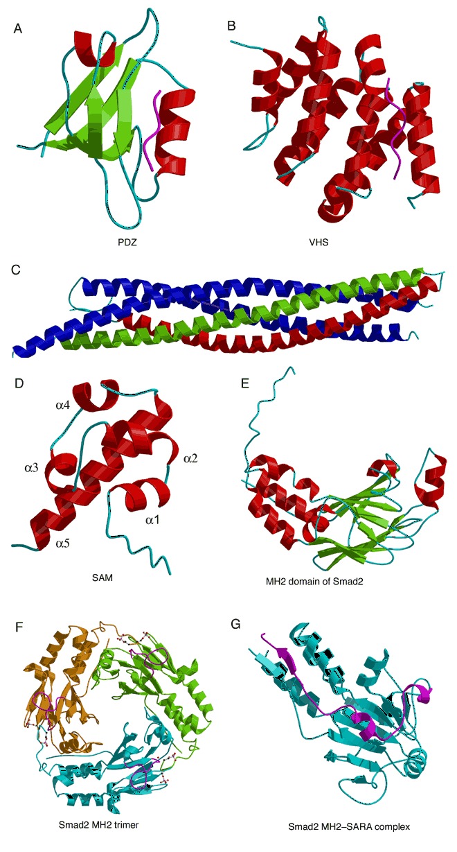
Protein interaction domains. Bound peptides are represented by magenta worms. (A) The syntrophin PDZ domain bound to the peptide GVKESLV (PDB entry 2pdz). (B) VHS domain of GGA1 complexed with cation‐independent mannose‐6‐phosphate receptor C‐terminal peptide (PDB entry 1jwg). (C) SNARE fusion complex containing syntaxin‐1A (green), synaptobrevin‐II (red), and SNAP‐25B (blue; PDB entry 1sfc). (D) SAM domain from human EPHB2 receptor (PDB entry 1b4f). (E) MH2 domain from human Smad2 (PDB entry 1khx). (F) Homotrimer of a phosphorylated MH2 domain from human Smad2 (PDB entry 1khx). Phosphorylated Ser465 and ‐467 are represented by ball‐and‐stick models. The phosphoserine binding loop L3 is shown in magenta. (G) Complex of the Smad2 MH2 domain (cyan) with the SARA complex (magenta; PDB entry 1dev).