
Figure 15.
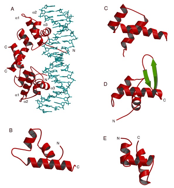
Variations of the helix‐turn‐helix (HTH) structural motif and the MADS box. The recognition helix is oriented horizontally in all but panel A. (A) Homodimer of the DNA‐binding domain of λ repressor bound to DNA (PDB entry 1lmb). The three helices of the HTH motif are labeled α1, α2, and α3 in each monomer. Both recognition helices (α3) sit in the major groove. (B) λ repressor (prokaryotic HTH; PDB entry 1lmb). (C) Engrailed homeodomain (PDB entry 1hdd). (D) Globular domain of histone H5 (PDB entry 1hst). This example of the winged helix motif has only one wing (the β hairpin). (E) Purine repressor (purR; PDB entry 1pru).